
Exams
Ultrasound or sonography utilizes sound waves to produce images of various organs or structures. Since there is no radiation, it is completely safe, even in early pregnancy.
At LSG Imaging, we perform many different types of ultrasound including, but not limited to, abdominal, pelvic, thyroid, scrotal, hernia detection and assessment, and musculoskeletal examinations. Ultrasound is also utilized to guide the radiologist when performing biopsies and cyst aspirations.
LSG Imaging is proud to have been the developers of transvaginal ultrasound. We, therefore, have special expertise in the field of GYN.
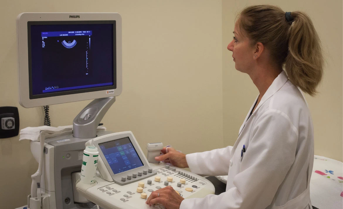
All ultrasound examinations are performed by the radiologist physician and not by a technologist. This, we believe, has great benefits to our patients. The patients are able to interact with the radiologist in detailing their history and their concerns. The examination can be “tailor made” to the patient. Also, because the radiologist is performing the examination, the results are generally communicated to the patient at the time of the study.
Hernia Ultrasound
Abdominal wall hernias are extremely common and often not diagnosed or misdiagnosed. Some of the more typical hernias include umbilical, inguinal and femoral (groin). Ultrasound is very effective in demonstrating their presence and is performed expertly and in a dynamic fashion. At our office, this exam is always performed by the Radiologist to optimize the examination.
Musculoskeletal Ultrasound
Many musculoskeletal conditions can be evaluated and diagnosed using ultrasound in our expert and competent fashion. For patients with cardiac pacemakers, this may be the only way to diagnose certain conditions. The most common condition we evaluate is related to the rotator cuff of the shoulder. Other conditions are also amenable.
Thyroid Ultrasound
Because it is noninvasive and involves no radiation, this examination is the most common exam used to evaluate the thyroid gland for the presence of masses, tumors and nodules. Should there be a need to biopsy any of the above, it is also used to help guide a needle to the target site.
Breast Ultrasound
A breast ultrasound is a diagnostic imaging procedure that uses high-frequency sound waves to produce images of the breast tissue and examine it for solid masses and other abnormalities. This procedure is often performed for patients with dense breasts on a mammogram. It is also performed when there is an abnormality in a mammogram and/or if a patient has a concern about a possible lump.
In addition to diagnosing masses and other abnormalities, breast ultrasound can also be used to:
- Determine the cause of pain, swelling and redness in the breast.
- Determine whether breast lumps are fluid-filled cysts or solid masses.
- Screen for breast cancer in patients with implants.
- Guide the placement of a needle to drain fluid or biopsy abnormal breast tissue.
Ultrasound: Frequently Asked Questions
Who performs my ultrasound at LSG Imaging, a doctor or a technologist?
At LSG Imaging, a board-certified radiologist performs every ultrasound personally. That is a meaningful distinction from most imaging centers, where a sonographer or technologist acquires the images and a radiologist reviews them later, often off-site.
At our Beverly Hills and Santa Monica locations, the physician interpreting your exam is in the room with you. You describe your history and concerns directly to the radiologist conducting the study. In most cases, you receive a verbal summary of preliminary findings before you leave. This approach is standard at LSG Imaging and has been for more than 40 years.
What is an ultrasound, and how does it work?
Ultrasound is a non-invasive imaging exam that uses high-frequency sound waves to produce real-time images of internal organs, tissues, and blood vessels. A small handheld device called a transducer is placed against the skin and emits sound waves that bounce off internal structures. A pelvic ultrasound usually is also performed with an intravaginal transducer. A computer converts those echoes into detailed images that your radiologist interprets during the exam.
Unlike CT or X-ray, ultrasound does not use ionizing radiation. It is widely used for evaluating soft tissue structures throughout the body, including the abdomen, pelvis, thyroid, breast, and musculoskeletal system.
How is ultrasound different from an MRI or CT scan?
Each imaging modality works differently and is suited to different clinical questions. Ultrasound uses sound waves, produces real-time images, involves no radiation, and is well suited for evaluating soft tissue organs, blood flow, and structures in the abdomen, pelvis, thyroid, and breast. It is also the preferred first-line imaging tool for pregnant patients and younger women because of its safety profile.
CT (computed tomography) uses X-ray technology to produce cross-sectional images of the body and is particularly effective for evaluating complex anatomy, detecting certain cancers, assessing vascular structures, and capturing a wide field of view quickly. CT does involve a dose of ionizing radiation and, for some exams, a contrast injection.
MRI (magnetic resonance imaging) uses magnetic fields and radio waves rather than radiation. It provides superior detail for soft tissues including the brain, spine, and joints, but takes longer, requires patients to lie still inside a tunnel-style scanner, and is not appropriate for patients with certain implanted metal devices.
Your referring physician selects the modality based on the specific clinical question. In many cases, ultrasound is ordered first because of its speed, safety, and real-time imaging capability. If your physician ordered an ultrasound and you are unsure why, our radiologists are happy to explain the reasoning during your appointment.
Is ultrasound safe, and does it use radiation?
Ultrasound does not use radiation of any kind. It relies entirely on sound waves, which makes it one of the safest imaging modalities available. There are no known risks associated with diagnostic ultrasound when performed by qualified imaging professionals.
Because it involves no radiation, ultrasound is the preferred imaging tool for pregnant patients, young women, and anyone who needs repeated imaging over time. It is also safe for pediatric patients and patients with implanted devices.
What conditions can an ultrasound detect or help diagnose?
Ultrasound is one of the most versatile tools in diagnostic radiology. It is used to evaluate a wide range of conditions across multiple organ systems. Common clinical applications include:
- Abdominal pain or abnormalities: Evaluation of the liver (fatty liver, masses, cirrhosis), gallbladder (gallstones, inflammation), spleen, kidneys (cysts, stones, obstruction), aorta for possible aneurism, pancreas, abdominal wall and inguinal hernias, and the appendix, just to mention the most common reasons.
- Pelvic conditions in women: Ovarian cysts, uterine fibroids, endometriosis evaluation, pelvic pain, and abnormal bleeding.
- Thyroid: Nodules, enlargement, or suspected masses in the thyroid gland
- Breast: Characterizing findings identified on mammography or physical exam, distinguishing cysts from solid masses.
- Vascular conditions: Deep vein thrombosis, carotid artery disease, and blood flow abnormalities.
- Musculoskeletal: Tendon tears, ligament injuries, joint effusions, and soft tissue masses
- Fertility evaluation: Pelvic ultrasound and ovarian follicle assessment for patients, working with reproductive specialists.
- Fetal development: Obstetric monitoring during pregnancy
Your referring physician will specify the clinical question being addressed. Our radiologists review findings in the context of your history and provide a complete diagnostic report.
Can an ultrasound detect cancer?
Ultrasound can identify masses, nodules, and structural abnormalities that may warrant further evaluation, including those that could represent cancer; many of those can or may undergo ultrasound guided needle biopsies. It is frequently used to evaluate breast lumps, thyroid nodules, ovarian masses, liver lesions, and kidney tumors. In many cases, ultrasound is the first imaging study that raises a flag prompting further workup.
However, ultrasound cannot confirm a cancer diagnosis on its own. Confirming malignancy requires tissue sampling, which is either a biopsy or surgical pathology. Ultrasound can also be used to guide biopsy procedures, helping physicians place a needle precisely into a suspicious lesion.
If your ultrasound identifies a finding that needs further evaluation, our radiologist will explain what was seen and outline recommended next steps clearly before you leave.
What is a transvaginal ultrasound, and is it uncomfortable?
A transvaginal ultrasound is a pelvic imaging exam in which a lubricated transducer is gently inserted into the vagina to produce detailed images of the uterus, ovaries, and surrounding pelvic structures. Because the transducer is positioned closer to the organs being evaluated, it produces clearer images than a transabdominal scan, particularly for smaller structures or early findings.
Most patients tolerate transvaginal ultrasound without significant discomfort. You may feel mild pressure during the exam, but it should not be painful. The procedure takes only a few minutes. No fasting nor special preparation is typically required beforehand. A full bladder is helpful initially for the transabdominal portion of the exam.
Our founders co-developed the technique of transvaginal ultrasound in 1984, a protocol now used by fertility and gynecological practices internationally.
At LSG Imaging, every pelvic ultrasound, whether transabdominal or transvaginal, is performed by a board-certified radiologist who will explain what is being evaluated and answer your questions throughout the exam.
How should I prepare for an ultrasound appointment?
Preparation depends on the type of ultrasound ordered. Specific instructions will be provided before your appointment, but general guidelines include:
- Abdominal ultrasound: Fasting for several hours beforehand is typically required to reduce bowel gas, optimize visualization of the gall bladder, and improve image quality.
- Pelvic ultrasound (transabdominal): A full bladder is usually needed. Drink water and avoid urinating for at least one hour before your appointment.
- Pelvic ultrasound (transvaginal): No special preparation is generally required.
- Thyroid, breast, or musculoskeletal ultrasound: No preparation is typically needed.
- Vascular ultrasound: Instructions vary depending on the vessels being examined.
Our care team sends preparation instructions in advance of every appointment. If you have any questions about your specific exam, call our Beverly Hills or Santa Monica office and our staff will walk you through the details.
How long does an ultrasound exam usually take?
Most ultrasound exams at LSG Imaging are completed within 15 to 45 minutes, depending on the area being evaluated and the clinical complexity of the study. Simpler exams, such as thyroid or superficial soft tissue studies, typically take less time. Pelvic, abdominal, and fertility ultrasounds may take longer when multiple structures need to be assessed in detail.
Because our radiologists perform every exam personally and review findings in real time, the overall appointment is efficient and you typically leave with a clear understanding of your results.
Is an ultrasound painful or uncomfortable?
Ultrasound is generally painless. The transducer is pressed gently against the skin, and a small amount of warm gel is applied to improve contact and image quality. Most patients find the experience entirely comfortable.
In some cases, mild discomfort is possible. Pelvic or abdominal ultrasounds can cause brief pressure if your bladder is full. Transvaginal ultrasound involves placement of an internal transducer. If you have any concerns before or during your exam, let our team know and we will do our best to keep you comfortable throughout.
Do I need a referral for an ultrasound?
Referral requirements vary by exam type. Many diagnostic ultrasound exams require a physician referral. However, some screening ultrasounds, including certain pelvic or fertility-related studies, may be scheduled without a referral depending on your situation.
When you call our Beverly Hills or Santa Monica office to schedule, our staff will confirm whether your specific exam requires a referral, verify any insurance questions, and walk you through the next steps. Our goal is to make scheduling as straightforward as possible.
When will I receive my ultrasound results?
At LSG Imaging, our radiologists perform every ultrasound personally and review findings in real time during your exam. In most cases, you will receive a verbal summary of your preliminary results before you leave. A formal written report is sent to your referring physician within 24 to 48 hours.
This is a meaningful distinction from most imaging centers, where a technologist performs the scan and a separate radiologist reviews it later off-site. Our direct approach means you are not waiting days for even an initial understanding of what the exam showed.
Why choose LSG Imaging for ultrasound in Beverly Hills or Santa Monica?
LSG Imaging has served the greater Los Angeles area for more than 40 years. Our approach to patient care differs from your typical experience at hospital-based imaging departments or large imaging chains:
- Radiologist-performed exams: Every ultrasound at LSG Imaging is performed personally by one of our board-certified radiologists. You speak directly with the physician reading your images, not a technologist whose work is sent elsewhere for interpretation.
- Same-day results in most cases: Because our radiologists review findings in real time, you leave with a clear understanding of what your exam showed.
- Four decades of subspecialty expertise: Our radiologists bring focused experience in women’s imaging, fertility, abdominal, and musculoskeletal ultrasound.
- Two convenient Los Angeles locations: Our Beverly Hills and Santa Monica offices offer short wait times, flexible scheduling, and a comfortable outpatient setting.
- Established clinical history: LSG Imaging was the first center in California to offer FDA-approved wide-angle 3D mammography, and our founders co-developed the technique of transvaginal ultrasound in 1984, a protocol now used by fertility and gynecological practices internationally.
Patients and referring physicians across West Los Angeles, Brentwood, Century City, Culver City, and surrounding communities have trusted LSG Imaging for consistent, high-quality diagnostic imaging for decades.